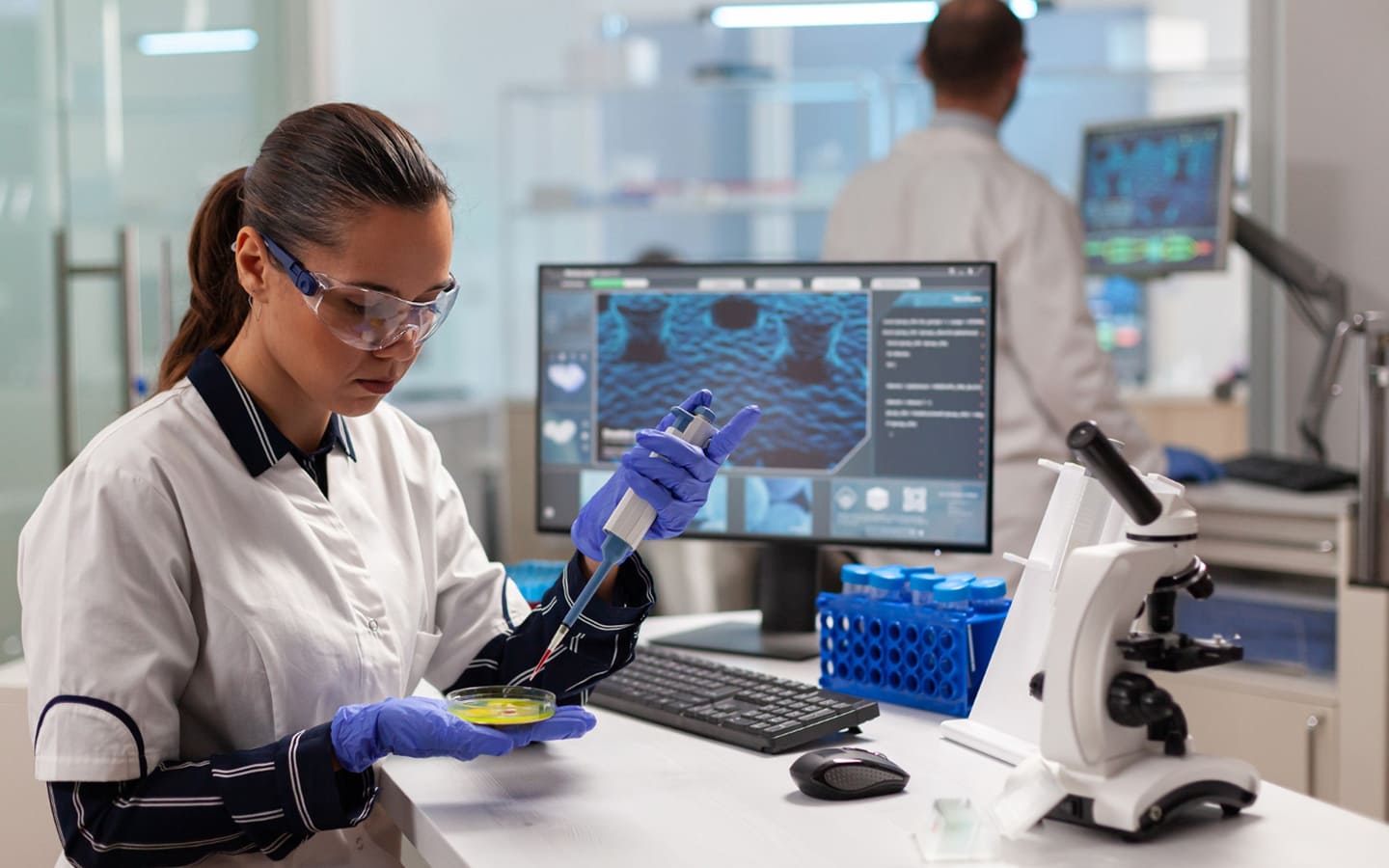
CW-EPR spectroscopy provides a highly sensitive and specific method for probing the local structure, dynamics, and environment of proteins (Garcia-Rubio, 2020) and lipid assemblies (Guzzi and Bartucci, 2015). The Site-Directed Spin Labeling Electron Paramagnetic Resonance (SDSL-EPR) spectroscopy offers a unique approach to elucidate the structure and dynamics of biomolecules in solution.
The approach combines protein chemistry/molecular genetics to target spin probes into biomolecules (most commonly peptides and proteins) with EPR analysis using modifications that can accommodate small biological samples under a variety of conditions (pure protein, tissue, live cells, etc.).
- Consultation on sample conditions and spectral acquisition parameters
- Protein chemistry if spin-labeling is required.
- Labeling with lipid spin probes for nanoparticle samples composed of lipid assemblies (liposomes, NLP/nanodisc/emulsions)
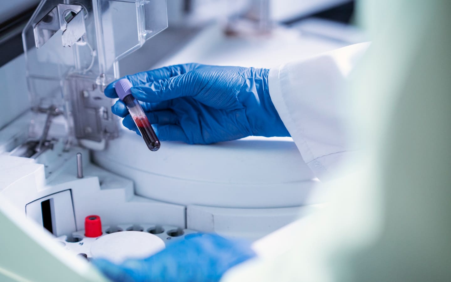
- Sample preparation, including sample concentration.
- Recording of EPR spectra with optimal parameters and temperatures (if temperature-dependent studies are needed).
- For probe accessibility/environment measurements, power saturation measurements are collected in the presence of distinct relaxation agents.
- Collection and plotting of data
- Computational analysis of line widths, amplitudes, and fitting of power saturation curves
- Written report of results
The EPR instrument services are categorized under three different service types, depending on the mode of data collection.
- Simple room temperature (RT) scan.
- Simple low temperature (LT) scan. Service also includes flash freezing of samples.
- EPR power saturation scanning.